


Optical Microscopy Application: Fluorescence
Authors: Stephan Briggs
Fluorescence microscopy is an optical microscopy technique that utilizes fluorescence, which is induced using fluorophores, as opposed to absorption, scatter, or reflection. A fluorophore is a type of fluorescent dye used to mark proteins, tissues, and cells with a fluorescent label for examination by fluorescence microscopy. A fluorophore works by absorbing energy of a specific wavelength region, commonly referred to as the excitation range, and re-emitting that energy in another specific wavelength region, commonly referred to as the emission range.
Fluorescence microscope systems can range from very simple, such as an epifluorescent microscope, to extremely complex, such as confocal or multiphoton systems. Whether simple or complex, fluorescence microscopes share the same basic concept: excitation energy is used to illuminate a sample which then emits a type of wavelength energy, albeit weak, that is quantifiable. The excitation and emission wavelengths do not share the same center wavelength, and this allows specialized optical filters to increase overall contrast and signal.
Image Appearance
Figure 1 shows a real-world fluorescence sample. The sample is excited by a shorter wavelength source (350 - 500nm), and then emits a fluorescent wavelength that is longer than the excitation wavelength (500nm +). Images such as this cannot be captured if not for the advanced optical filtering techniques in fluorescence microscopy which allow narrow bandwidths of light to propagate through to the sensor resulting in very crisp, high contrast images. Fluorescent proteins in specimens cause the unique emission colors. These proteins, such as GFP, are often derived from marine life.
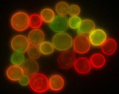
Figure 1: Fluorescence Image of Microspheres
Technical Details
In general, the technical details of a fluorescence microscope mirror any standard brightfield illumination microscope or darkfield illumination microscope. The innovation comes from the intricate filtering techniques that are used to selectively utilize narrow bands of light. The three critical filters needed for a precision fluorescence microscope are the excitation, dichroic, and emission filters. For more in-depth information, please read Fluorophores and Filters in Fluorescence Microscopy.
- Excitation Filter: placed within the illumination path of a fluorescence microscope. It filters out all wavelengths of the light source except for the excitation range of the fluorophore or specimen under inspection.
- Dichroic Filter: placed between the excitation filter and emission filter at a 45° angle. It reflects the excitation signal towards the fluorophore under inspection and transmits the emission signal towards the detector.
- Emission Filter: placed within the imaging path of a fluorescence microscope. It filters out the entire excitation range of the fluorophore under inspection and transmits the emission range of the fluorophore.
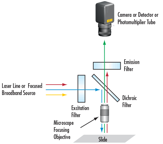
Figure 2: Basic Optical Filtering Arrangement for Fluorescence Microscopy
Additional optical microscopy applications include brightfield illumination, darkfield illumination, phase contrast, and differential interference contrast.


or view regional numbers
QUOTE TOOL
enter stock numbers to begin
Copyright 2023, Edmund Optics Inc., 101 East Gloucester Pike, Barrington, NJ 08007-1380 USA
California Consumer Privacy Acts (CCPA): Do Not Sell or Share My Personal Information
California Transparency in Supply Chains Act
This content may include material that has been generated or modified using artificial intelligence (AI).
The FUTURE Depends On Optics®