Engineers are available to assist.


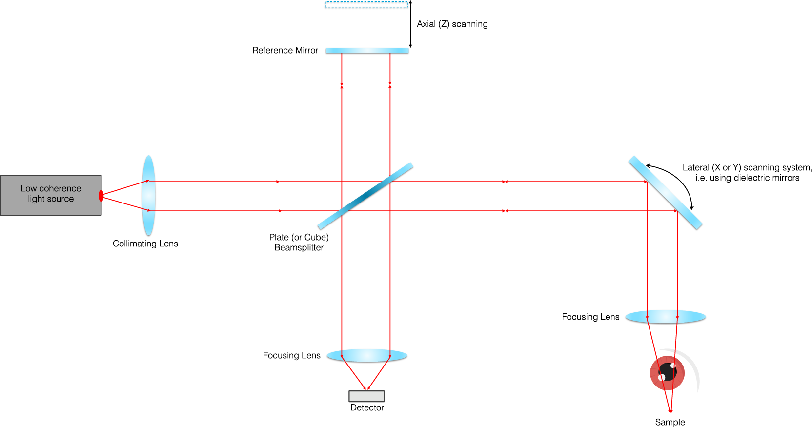
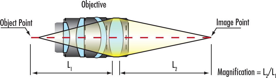
Optical coherence tomography (OCT) is a non-invasive, high-resolution optical imaging technology that creates cross-sectional images from interference signals received from an object under investigation and a reference optic. OCT is commonly used in the medical field to obtain real-time, two-dimensional (2D) and three-dimensional (3D) images in-vivo for direct visualization of tissue structures.
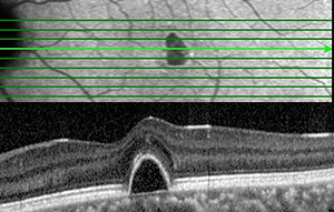
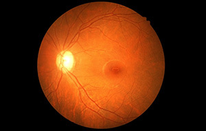
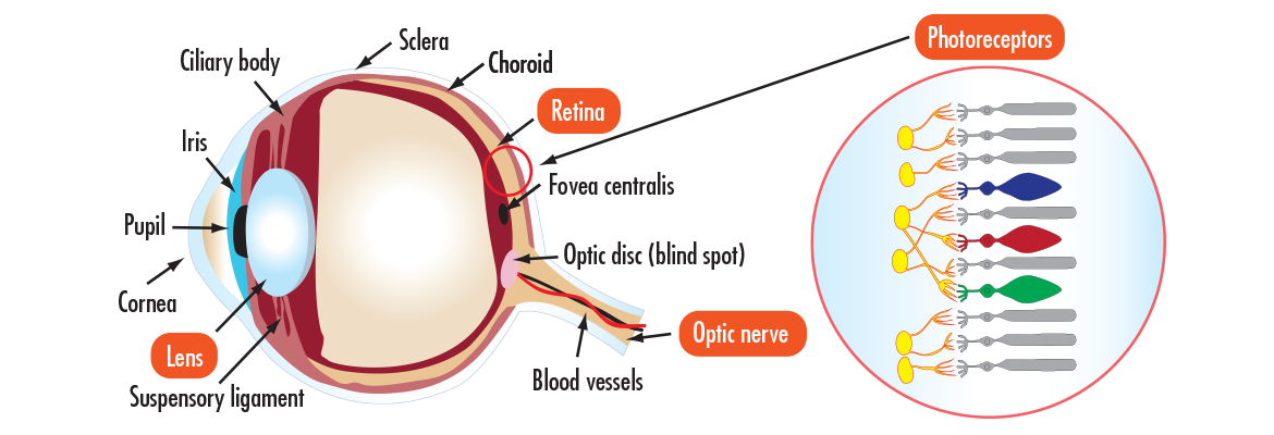
OCT allows improved diagnosis of ophthalmic diseases, such as Age-related Macular Degeneration, AMD (a disease of the retina), which causes blurred vision or diabetic retinopathy, by quantitatively characterizing changes in the structure and appearance of retinal tissue. The effectiveness of therapies can also be tracked using OCT by quantifying the retinal thickness and biomarkers to determine if the disease is progressing. OCT can also be adapted for cardiology, where it enables the diagnosis of the likelihood of a heart attack. One of the leading causes for heart attacks is atherosclerosis which occurs when ruptured fatty plaques and calcium build-up inside the lining of the artery wall, blocking blood flow. OCT allows the detection of vulnerable plaques prior to rupture by visualizing plaques in the arterial wall with an image resolution of 5-7 µm to determine the size, shape, and location of the plaque. In both ophthalmology and cardiology, the ability to accurately diagnose degenerative diseases early makes treatments more effective and provides a better understanding of cellular progression and pathways of many incurable diseases.
Edmund Optics supplies a wide range of optics ideal for OCT systems, including plate and cube beamsplitters, broadband dielectric mirrors, lenses, illumination sources and whole Lumedica benchtop OCT Imaging Systems.
Optical coherence tomography (OCT) is a powerful medical imaging technique which utilizes light to capture high resolution, three dimensional images from optical scatter in biological tissue. The principles are based off of simple interferometry with near infrared light to effectively penetrate the biological medium. There is a tradeoff to depth penetration and resolution that other technologies can address, but OCT is often coupled with these to ensure accuracy and multi model images.
There are a variety of advanced diagnostic techniques used to treat diseases and ailments of the eye. More and more techniques and devices are emerging due to the progression of optical technology, increasing the accuracy and timeliness of which ailments are diagnosed and providing a pathway for understanding and finding cures for incurable diseases.
Laser eye surgery intended to correct human vision. LASIK is a refractive surgery to correct myopia and astigmatism by using a laser to reshape the cornea of the eye, therefore improving visual acuity.
Branch of medicine that deals with the anatomy, study, and diseases of the eye. The eye is one of the leading indicators for diagnosing a number of serious ailments. Due to the eyes ease of access and high level of transmission, it has become the “gold standard” for non-invasive medical imaging through various means and technologies such as optical coherence tomography (OCT).
Automated biometric identification using mathematical algorithms to identify and properly recognize an individual’s iris/pupil. This form of biometric recognition is very reliable, as a human’s eye pattern is completely unique, stable over long periods of time, and can be distinguished and recognized over a great distance.

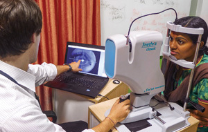
Forus Health leveraged Edmund Optics’ optical design expertise and off-the-shelf components to build 3nethra, an affordable and portable ophthalmic device for diagnosing preventable eye diseases in India. India has a low ratio of ophthalmologists to population, and this easy-to-use device helps identify and prevent cataracts and glaucoma, along with diabetic retinopathy, refractive errors, and cornea problems.
Learn More
Below are common ailments of the eye that are detected by advanced diagnostic technology. These ailments occur for a variety of reasons such as age or blunt trauma, but are more easily detected and treated due to optical advancements within the industry, which provides a means for updating medical technology and equipment for quick, portable, and simplified use.


or view regional numbers
QUOTE TOOL
enter stock numbers to begin
Copyright 2023, Edmund Optics Inc., 101 East Gloucester Pike, Barrington, NJ 08007-1380 USA
California Consumer Privacy Acts (CCPA): Do Not Sell or Share My Personal Information
California Transparency in Supply Chains Act
This content may include material that has been generated or modified using artificial intelligence (AI).
The FUTURE Depends On Optics®