Engineers are available to assist.

|
|
Network of users collaborating to develop and improve technology |
|
|
Cost-effective microscopy systems available to a broad audience |
|
|
Microscope systems that are simple to build and modify |
|
|
Facilitates spread of life-saving advanced diagnostic techniques |
Open-source technology is based around a community of users collaborating to develop, test, share, and improve the technology. Technical information is made available and the users of the technology share ideas and developments related to the product. The most commonly open-sourced product is software as it is well suited for editing and manipulation by a network of people. However, other technologies such as microscopy can be open-sourced as well, making otherwise very cost-prohibitive products available to a much broader audience. Open-sourced DIY microscopy provides an accessible, cost-effective, and flexible way for microscopy systems to be used for research and prototyping.
For neuroscience researchers, studying neural activity on freely behaving animals is critical for research, but traditional microscopes used to study animal brains tend to be cumbersome and expensive, and must be stationary when used. This makes them extremely inconvenient for observing the neurons of a mouse exploring its environment or interacting with other animals. The Khakh, Silva, and Golshani labs at the University of California, Los Angeles (UCLA) created an open-source, head-mounted miniature fluorescent microscope to study neural activity on freely behaving mice. Any neuroscience lab can build this optical device at a fraction of the cost of a commercial fluorescence microscope.
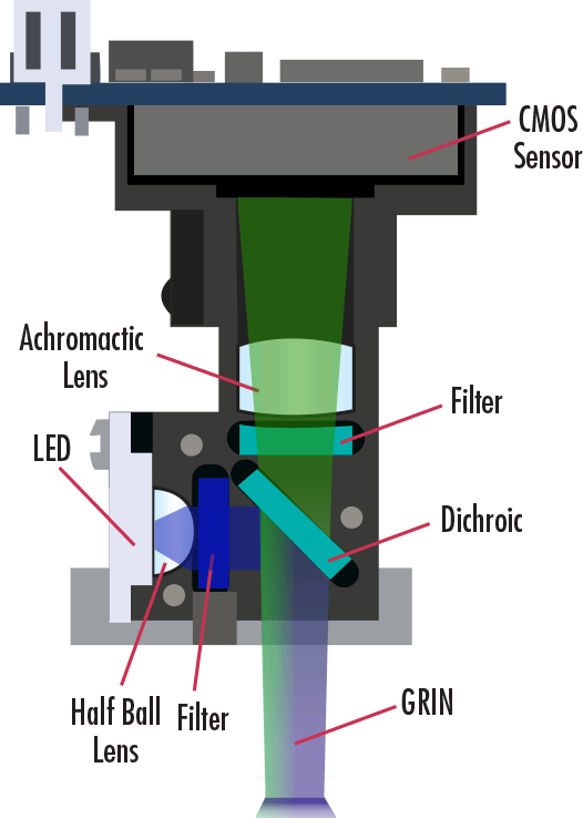
The Miniscope mounts onto the head of a mouse to image neurons firing and identify the differences in neural activity between healthy brains and those with neurological conditions. True to their goal of being open-source, the UCLA research team created the design for this microscope and posted the parts list, instructions for building and using the Miniscope, and links to video tutorials on a custom wiki website. This wiki provides a centralized location for sharing design files, source code, and other relevant information so that a community of users and scientists from around the world can share ideas and developments related to this important imaging technique. The central location of information helps spread the technology to the larger neuroscience community, establishing a foundation of users that will continue advancing this technology and contribute back to the project.
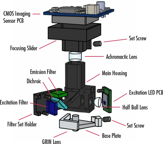
This DIY microscope includes off-the-shelf components from Edmund Optics® including achromatic lenses, half-ball lenses, and GRIN lenses. The Miniscope represents the building block of a stable platform for labs to add components to in order to support their specific usage of the microscope. Over 300 labs are using this Miniscope today, many of which have customized the device to their unique research needs. To learn more or purchase the complete set of components for the Miniscope, visit this webpage.
 Where can I can purchase the complete set of components for the Miniscope?
To learn more about the Miniscope and to purchase the complete set of components, visit this webpage.
How much does the Miniscope weigh?
The completely assembled Miniscope weighs 3 grams. This low mass allows it to be mounted on the head of freely behaving mice.
Where can I find all of the design files, source code, and other relevant information about the Miniscope?
Is the Miniscope project still under development?


or view regional numbers
QUOTE TOOL
enter stock numbers to begin
Copyright 2023, Edmund Optics Inc., 101 East Gloucester Pike, Barrington, NJ 08007-1380 USA
California Consumer Privacy Acts (CCPA): Do Not Sell or Share My Personal Information
California Transparency in Supply Chains Act
This content may include material that has been generated or modified using artificial intelligence (AI).
The FUTURE Depends On Optics®